

A lip ulcer is a loss of the epithelial surface of the lip, resulting in an open sore that exposes the underlying tissue. It presents as a painful or painless lesion with a central area of tissue loss, often covered by a gray-white or yellowish fibrin layer surrounded by an erythematous border.
Cheilitis is defined as a superficial inflammatory condition affecting the lip (vermillion border), and can present as erythema, scaling, fissuring, or crusting. It may be generalized, or localized to one or more lesions. Although some edema may be present, the main manifestation is discomfort. For information on lip swelling with little or no discomfort see Lip swelling.
(See also Evaluation of the Dental Patient.)
Lip ulcers
A number of infectious, neoplastic, inflammatory or autoimmune, medication-related disorders can cause lip ulcers:
Herpes labialis (herpes simplex virus infection): A small cluster of fluid-filled vesicles that rupture to form an ulcer on the lip's vermilion border; commonly referred to as a cold sore or a fever blister.
Erythema multiforme: Multiple bullae that rupture quickly and leave crusting hemorrhagic ulcers on labial mucosa (1). This ulcerative mucocutaneous condition is an immune reaction usually triggered by herpes simplex virus. Erythema multiforme has a variety of appearances and often causes painful oral mucositis.
Primary syphilis (chancre): A painless ulcer with hard edges. Oral chancres are commonly seen on lips (upper lip more common in males; lower lip more common in females) (2).
Recurrent aphthous stomatitis: Major aphthous ulcers may occur on the lips (labial mucosa) and may persist for weeks to months. Multiple recurrent smaller painful ulcers also typically occur in other parts of the oral mucosa.
Drug-induced ulcers: Lip ulcers can result from direct cytotoxic effects (chemotherapy, alendronate) or hypersensitivity reactions (eg, Drug-induced ulcers: Lip ulcers can result from direct cytotoxic effects (chemotherapy, alendronate) or hypersensitivity reactions (eg,Stevens-Johnson syndrome, drug reaction with eosinophilia and systemic symptoms [DRESS]).
Systemic inflammatory or autoimmune conditions: Systemic diseases that are associated with aphthous stomatitis that can involve the vermillion border include celiac disease, Behçet disease, inflammatory bowel diseases, and PFAPA syndrome (periodic fever, aphthous stomatitis, pharyngitis, and adenitis). Autoimmune bullous diseases such as pemphigus vulgaris and bullous pemphigoid can also present with lip ulcerations through immune-mediated mechanisms.
Trauma: Traumatic ulcers can result from mechanical injury such as accidental biting, sharp tooth edges, dental appliances, or external trauma.
Other conditions that can present with lip lesions and may mimic ulcers include:
Verruca vulgaris (common wart): Pebbly surfaced, painless growth. This benign condition can spread by autoinoculation.
Actinic cheilitis: Irregular pale, red, or variably colored dry and scaly precancerous growths affecting the lips. This common premalignant condition is caused by chronic exposure to ultraviolet light.
Erythroplakia or leukoplakia: Red or white patches that typically develop on the floor of the mouth or lateral tongue, and buccal mucosa but can rarely affect the lips (3). These patches may be associated with dysplasia and squamous cell carcinoma.
Oral squamous cell carcinoma: May present variably as a hyperkeratotic nodule or plaque, ulcer with hard edges.
Peutz-Jeghers syndrome: Autosomal dominant condition that includes benign hyperpigmented (dark blue, brown, black) macules of skin and mucosa (especially lip and buccal mucosae), gastrointestinal hamartomatous polyps, and (unrelated to the macules and polyps) predisposition to various cancers.


This photo is a close-up of a herpes simplex virus type 1 (HSV-1) lesion on the lower lip that shows clusters of vesicles on an erythematous base.
This photo is a close-up of a herpes simplex virus type 1 (HSV-1) lesion on the lower lip that shows clusters of vesicl
CDC


This photo shows oral and palmar lesions.
This photo shows oral and palmar lesions.
Photo courtesy of Karen McKoy, MD.


Syphilitic chancres may appear on or around the mouth.
Syphilitic chancres may appear on or around the mouth.
CDC


© Springer Science+Business Media
This photo is a close-up of a herpes simplex virus type 1 (HSV-1) lesion on the lower lip that shows clusters of vesicles on an erythematous base.
This photo is a close-up of a herpes simplex virus type 1 (HSV-1) lesion on the lower lip that shows clusters of vesicl
CDC
This photo shows oral and palmar lesions.
This photo shows oral and palmar lesions.
Photo courtesy of Karen McKoy, MD.
Syphilitic chancres may appear on or around the mouth.
Syphilitic chancres may appear on or around the mouth.
CDC
© Springer Science+Business Media
Lip ulcers references
1. Ayangco L, Rogers RS 3rd. Oral manifestations of erythema multiforme. Dermatol Clin. 2003;21(1):195-205. doi:10.1016/s0733-8635(02)00062-1
2. Zhou X, Wu MZ, Jiang TT, Chen XS. Oral Manifestations of Early Syphilis in Adults: A Systematic Review of Case Reports and Series. Sex Transm Dis. 2021;48(12):e209-e214. doi:10.1097/OLQ.0000000000001538
3. de Azevedo AB, Dos Santos TCRB, Lopes MA, et al. Oral leukoplakia, leukoerythroplakia, erythroplakia and actinic cheilitis: Analysis of 953 patients focusing on oral epithelial dysplasia. J Oral Pathol Med. 2021;50(8):829-840. doi:10.1111/jop.13183
Cheilitis (Lip Inflammation)
Cheilitis is acute or chronic inflammation of the lips. It may be caused by infection, sun damage, medications or irritants, allergy, or underlying disease. Inflammation primarily affects the vermilion and vermilion border. Swelling, redness, and pain of the lips occurs; other changes may include cracks, fissures, erosions, crusts, and scale.
Common causes of cheilitis include:
Angular cheilitis: Inflammation, crusting, painful fissures, and maceration in the corners of the mouth.
Actinic cheilitis: Sun damage causing thin, atrophic mucosa with erosions; predisposes to malignancy
Eczematous cheilitis: Red, dry lips (sometimes termed chapped lips) typically caused by contact irritants or sometimes by allergens or as part of atopic dermatitis
Rare types of cheilitis include cheilitis glandularis, cheilitis granulomatosa, and plasma cell cheilitis.
Children with Kawasaki disease may develop erythematous, dry, swollen, and cracked lips that can mimic cheilitis, along with strawberry tongue. Prompt diagnosis and treatment is required to prevent adverse outcomes.
Angular cheilitis
Angular cheilitis (angular stomatitis) is the most common form; inflammation, crusting, painful fissures, and often maceration develop in the corners of the mouth. Typical causes include:
Excessively worn teeth or dentures that do not adequately separate the jaws, creating skin folds at the corners of the mouth in which saliva accumulates
Candida species (or sometimes Staphylococcus aureus)
Iron deficiency, vitamin B complex deficiency (especially riboflavin, cobalamin)

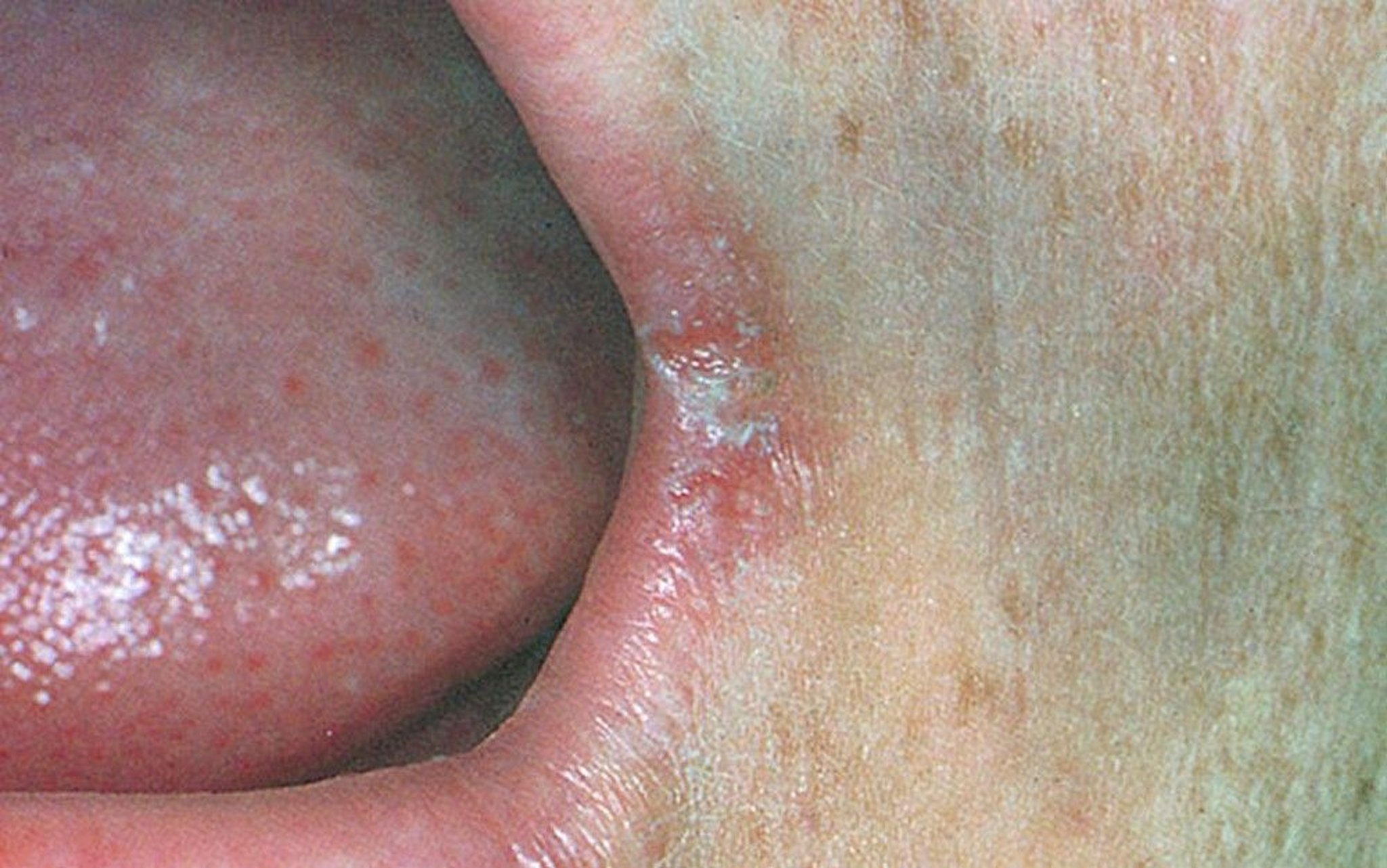
Angular cheilitis (perlèche) produces painful cracks at the corners of the mouth. Erythema and crusting at the labial commissure also is seen in this photo.
© Springer Science+Business Media
Treatment of angular cheilitis may involve replacing dentures or restoring proper tooth size with partial dentures, crowns, or implants, which helps reduce the folds at the corners of the mouth. Primary treatment of other causes may include the use of antifungals (eg, clotrimazole cream), antibiotics (eg, mupirocin ointment), or iron or vitamin B supplementation as appropriate.Treatment of angular cheilitis may involve replacing dentures or restoring proper tooth size with partial dentures, crowns, or implants, which helps reduce the folds at the corners of the mouth. Primary treatment of other causes may include the use of antifungals (eg, clotrimazole cream), antibiotics (eg, mupirocin ointment), or iron or vitamin B supplementation as appropriate.
Actinic cheilitis
Actinic cheilitis (solar cheilosis) is a premalignant condition of the lip caused by chronic sun exposure. It is considered the equivalent of actinic keratosis of sun-exposed skin. Damage to the lips typically include dryness, scaling, and loss of distinction between the vermillion border and the keratinized epithelium. Excessive sun exposure may also result in redness and ulcerations, which must be evaluated by a dentist or physician for possible biopsy.
Prevalence of actinic cheilitis is low in the general population, but can be significantly higher in groups who participate in frequent outdoor activities (1). The highest risk groups appear to be males over 50 years of age and those with lightly pigmented skin.
The lower lip is predominantly affected. These lip changes are considered as a potential precursor to the development of squamous cell carcinoma.
The diagnosis is primarily by history and physical examination, though biopsy is usually recommended for histopathologic confirmation and to exclude squamous cell carcinoma or other invasive malignant lesions (2).
Treatment is the same as that for actinic keratosis, which is discussed in detail separately (see Actinic Keratosis). Reduction of sun exposure through use of hats with wide brims and use of sunscreens is highly recommended.
Cheilitis references
1. Rodriguez-Archilla A, Irfan-Bhatti A. Risk factors for actinic cheilitis: A meta-analysis. J Dent Res Dent Clin Dent Prospects. 2021;15(4):285-289. doi:10.34172/joddd.2021.047
2. Seoane J, Warnakulasuriya S, Bagán JV, et al. Assembling a consensus on actinic cheilitis: A Delphi study. J Oral Pathol Med. 2021;50(10):962-970. doi:10.1111/jop.13200
Drug Information for the Topic


