

Amenorrhea (the absence of menstruation) can be primary or secondary.
Primary amenorrhea is failure of menses to occur by age 15 years in patients with normal growth and secondary sexual characteristics (1). In addition, absence of menarche and of any breast development by age 13 should prompt evaluation for delayed puberty with primary amenorrhea.
Secondary amenorrhea is the absence of menses for 3 months in patients with previously regular menstrual cycles or for ≥ 6 months in patients with previously irregular menses (2).
General references
1. Yatsenko SA, Witchel SF, Gordon CM. Primary Amenorrhea and Premature Ovarian Insufficiency. Endocrinol Metab Clin North Am. 2024;53(2):293-305. doi:10.1016/j.ecl.2024.01.009
2. Gordon CM, Ackerman KE, Berga SL, et al. Functional hypothalamic amenorrhea: An Endocrine Society clinical practice guideline. Clin Endocrinol Metab 102 (5):1413–1439, 2017. doi: 10.1210/jc.2017-00131
Pathophysiology of Amenorrhea
Normally, the hypothalamus generates pulses of gonadotropin-releasing hormone (GnRH). GnRH stimulates the pituitary gland to produce gonadotropins, follicle-stimulating hormone (FSH), and luteinizing hormone (LH) (see figure ), which are released into the bloodstream. Gonadotropins stimulate the ovaries to produce estrogens (mainly estradiol), androgens (mainly testosterone), and progesterone. These hormones do the following:
Follicle-stimulating hormone activates aromatase in ovarian granulosa cells that surround the developing oocytes to convert androgens to estradiol.
Luteinizing hormone surges during the menstrual cycle; this surge promotes maturation of the dominant oocyte, release of the oocyte, and formation of the corpus luteum (which produces progesterone).
Estrogen stimulates the endometrium (lining of the uterine cavity), causing it to proliferate.
Progesterone changes the endometrium into a secretory structure and prepares it for implantation of the fertilized ovum (endometrial decidualization).
If ovulation occurs and the oocyte is not fertilized, estrogen and progesterone production decrease, and the endometrium breaks down and is sloughed as menses. Menstruation occurs 14 days after ovulation in a 28-day menstrual cycle.
Normal Menstrual Cycle
This figure shows the idealized cyclic changes in pituitary gonadotropins, estradiol (E2), progesterone (P), and uterine endometrium during the normal menstrual cycle. |


When part of this system malfunctions, ovulatory dysfunction occurs; the cycle of gonadotropin-stimulated estrogen production and cyclic endometrial changes is disrupted, resulting in anovulation, and menstrual flow may not occur.
Etiology of Amenorrhea
Amenorrhea can be classified based on a number of different criteria, such as:
Type: primary or secondary
Etiology: gonadal dysgenesis, structural, or endocrinologic
Primary amenorrhea may be caused by gonadal dysgenesis, congenital reproductive tract anomalies, or endocrinologic abnormalities.
Secondary amenorrhea may be caused by acquired reproductive tract structural abnormalities that interfere with menstrual function or obstruct menstrual flow or endocrinologic abnormalities.
Ovulatory dysfunction is the most common cause of amenorrhea, particularly secondary amenorrhea.
Gonadal dysgenesis (abnormal gonadal development) due to gene or chromosome abnormalities includes:
Turner syndrome: Absence of all or part of 1 of the 2 X chromosomes
46,XY gonadal dysgenesis (Swyer syndrome): Patients are genotypically male, but phenotypically female
Fragile X syndrome: An abnormality of the FMR1 gene on the X chromosome
Patients with these disorders often have incompletely formed gonads (streak gonads) and primary ovarian insufficiency (premature ovarian failure), resulting in primary amenorrhea and infertility. Genetic disorders that confer a Y chromosome increase the risk of ovarian germ cell cancer.
Structural causes of amenorrhea include:
Congenital reproductive tract anomalies (eg, vaginal septum, vaginal agenesis [most commonly due to mullerian agenesis, or Mayer-Rokitansky-Küster-Hauser syndrome], imperforate hymen)
Acquired structural abnormalities (eg, intrauterine adhesions [Asherman syndrome], cervical stenosis)
There are many potential endocrinologic causes. Common endocrinologic causes include:
Constitutional delay of puberty
Hyperprolactinemia (eg, due to pituitary adenoma, lactational amenorrhea during breastfeeding, use of antipsychotics)
Functional hypothalamic amenorrhea (eg, due to excessive exercise, eating disorders, disordered eating, stress [1, 2])
Primary ovarian insufficiency (POI) (eg, due to autoimmune disease, chemotherapy, or radiation therapy)
Pregnancy (most common cause in women of reproductive age)
Hormonal medications (eg, oral contraceptives, depot medroxyprogesterone)Hormonal medications (eg, oral contraceptives, depot medroxyprogesterone)
Progestin-only contraceptives (pills, injections, implants, and intrauterine devices) often cause irregular menses or amenorrhea. Combined estrogen/progestin contraceptives can cause amenorrhea if they are used continuously (placebo pills are skipped and hormone pills are taken every day) or for a long time (due to atrophic endometrium).
Less common endocrinologic causes include receptor or enzyme disorders (eg, complete androgen insensitivity syndrome, 5-alpha-reductase deficiency).
Amenorrhea due to ovulatory dysfunction
Amenorrhea due to ovulatory dysfunction is usually secondary but may be primary if ovulation never begins—eg, because of gonadal dysgenesis or due to an endocrinologic disorder. Patients with primary amenorrhea due to ovulatory dysfunction usually present with delayed puberty.
The most common causes of ovulatory dysfunction involve a disruption of the hypothalamic-pituitary-ovarian axis (See table ). Thus, causes include:
Perimenarche or perimenopause
Androgen excess (particularly due to polycystic ovary syndrome)
Pituitary dysfunction (eg, hyperprolactinemia cause by pituitary adenoma, pituitary irradiation),
Idiopathic (sometimes occurring when gonadotropin levels are normal)
Hypothalamic dysfunction may result in decreased GnRH production, which in turn may cause decreased gonadotropin production. A common cause is insufficient energy intake due to dietary restriction, undernutrition, or strenuous exercise or emotional stress. Women with amenorrhea due to hypothalamic dysfunction have lower levels of serum leptin (an anorectic hormone produced by fat cells); lower levels may contribute to decreased gonadotropin production (3).
The Central Nervous System-Hypothalamic-Pituitary-Gonadal Target Organ Axis
Ovarian hormones have direct and indirect effects on other tissues (eg, bone, skin, muscle). FSH = follicle-stimulating hormone; GnRH = gonadotropin-releasing hormone; LH = luteinizing hormone. |


Some Etiologies of Ovulatory Dysfunction
Etiology | Examples |
|---|---|
Hypothalamic dysfunction, functional | Inadequate calorie intake due to dieting, orthorexia*, eating disorders (eg, anorexia nervosa, bulimia), undernutrition (eg, due to food insecurity), restricted eating*, disordered eating*, or malabsorption Excessive calorie expenditure (if calories used exceed calorie intake) due to exercise or hypermetabolic states Metabolic disturbances (eg, associated with obesity, aging) Emotional stress Psychiatric disorders (eg, depression, obsessive-compulsive disorder, schizophrenia) Chronic disease, particularly gastrointestinal, renal, or hepatic (eg, Crohn disease, celiac disease, chronic kidney disease requiring hemodialysis, cirrhosis, sickle cell disease, cystic fibrosis, thalassemia major, or seizure disorders) Some medications, including psychoactive medications (eg, antipsychotics, antidepressants) and antiepileptics Misuse of some substances (eg, alcohol, opioids) Infections (eg, HIV infection, encephalitis, syphilis) |
Hypothalamic dysfunction, genetic or structural | Genetic disorders (eg, idiopathic gonadotropin-releasing hormone deficiency [also referred to as idiopathic hypogonadotropic hypogonadism or Kallman syndrome], monogenic obesity syndromes [eg, Prader-Willi syndrome], congenital hypopituitarism) Infiltrative inflammatory disorders of the hypothalamus (eg, Langerhans cell histiocytosis, sarcoidosis, tuberculosis) Neoplasms of the hypothalamus (eg, lymphoma, craniopharyngiomas, gliomas, metastatic disease) Irradiation to the hypothalamus |
Pituitary dysfunction | Tumors of the pituitary (eg, microadenoma, metastatic carcinoma, endodermal sinus tumor, other pituitary tumors that secrete hormones [eg, ACTH, thyroid-stimulating hormone, growth hormone, FSH, LH]) Other brain tumors (eg, meningioma, glioma, ependymoma) Irradiation of the pituitary Space-occupying lesions (eg, empty sella turcica, cerebral arterial aneurysm) Infiltrative disorders of the pituitary (eg, hemochromatosis, Langerhans cell granulomatosis, sarcoidosis, tuberculosis) Hyperprolactinemia (primary or secondary to other endocrinologic disorders [eg, thyroid disease], medications, or other substances) Genetic or developmental disorders (eg, idiopathic gonadotropin-releasing hormone deficiency, hypoplastic pituitary gland) Postpartum pituitary infarction and necrosis (Sheehan syndrome) Traumatic brain injury |
Ovarian dysfunction | Autoimmune disorders (eg, autoimmune oophoritis associated with myasthenia gravis, thyroiditis, or vitiligo) Gonadal dysgenesis due to genetic abnormalities, including chromosomal abnormalities (eg, congenital thymic aplasia, Fragile X syndrome, Turner syndrome [45,X], idiopathic accelerated ovarian follicular atresia) Metabolic disorders (eg, Addison disease, diabetes mellitus, galactosemia [†]) Viral infections (eg, mumps) Chemotherapy (eg, high-dose alkylating agents) Irradiation to the pelvis Ovarian tumors (eg, granulosa-theca cell tumors, Brenner tumors, teratomas, mucinous or serous cystadenomas, Krukenberg tumors, metastatic carcinoma) |
Other endocrine dysfunction | Androgen insensitivity syndrome (testicular feminization) Congenital adrenal hyperandrogenism (congenital adrenal hyperplasia—eg, due to 17-hydroxylase deficiency or 17,20-lyase deficiency) or adult-onset adrenal hyperandrogenism§ Ovotesticular difference of sexual development (formerly called true hermaphroditism)§ Tumors producing androgens (usually ovarian or adrenal)‡ Drug-induced virilization (eg, by androgens, antidepressants, danazol, or high-dose progestins)§Drug-induced virilization (eg, by androgens, antidepressants, danazol, or high-dose progestins)§ Obesity (which causes excess extraglandular production of estrogen) Tumors producing estrogens or tumors producing human chorionic gonadotropin (gestational trophoblastic disease) |
* Orthorexia is characterized by obsession with eating healthy foods, with associated restrictive behaviors that may result in restriction of nutrients and calories leading to amenorrhea. Restricted eating is a conscious effort to limit food intake, often driven by the desire to lose weight or control eating habits. Disordered eating describes unhealthy eating behaviors that do not meet the criteria for a specific eating disorder diagnosis. † Berry GT. Galactosemia and amenorrhea in the adolescent. Ann N Y Acad Sci. 2008;1135:112-117. doi:10.1196/annals.1429.038 ‡ Krassas GE. Thyroid disease and female reproduction. Fertil Steril. 2000;74(6):1063-1070. doi:10.1016/s0015-0282(00)01589-2 § Females with these disorders may have virilization or ambiguous genitals. | |
ACTH = adrenocorticotropic hormone; FSH = follicle-stimulating hormone; GnRH = gonadotropin-releasing hormone; LH = luteinizing hormone. | |
Amenorrhea due to structural reproductive tract abnormalities
In patients with anatomic or other structural abnormalities of the reproductive tract, menses are absent because either:
Menstrual blood is not produced: uterus is absent (due to congenital agenesis of the uterus) or the endometrium is not functioning (eg, due to intrauterine adhesions)
Menstrual flow is blocked: uterus, cervix, or vagina are blocked (due to congenital anomalies or acquired blockage [eg, cervical stenosis])
Mullerian agenesis (Mayer-Rokitansky-Küster-Hauser syndrome) is a syndrome that involves congenital anomalies of the female reproductive tract, sometimes with associated urinary tract abnormalities (4). It is the most common cause of primary amenorrhea, occurring in approximately 1 in 5000 females (5). Primary amenorrhea caused by obstructive abnormalities is usually accompanied by normal hormonal function, thus, external genital organs and other secondary sexual characteristics develop normally.
Likewise, patients with secondary amenorrhea due to structural abnormalities usually have normal reproductive endocrine function.
Some Causes of Amenorrhea due to Structural Reproductive Tract Abnormalities
Cause | Examples |
|---|---|
Congenital female genital anomalies | Vaginal, cervical, or uterine agenesis or aplasia (eg, müllerian agenesis) Imperforate hymen Pseudohermaphroditism Transverse vaginal septum |
Acquired uterine abnormalities | Asherman syndrome (intrauterine adhesions), usually caused by infection or an intrauterine procedure Endometrial ablation procedure Cervical stenosis Endometrial tuberculosis |
Obstruction results in accumulation of blood within the genital tract, which can cause the following complications:
Hematometra (accumulation of blood in the uterus), which can cause pelvic pain and uterine distention may be noted as a pelvic mass or bulging of the cervix
Hematocolpos (accumulation of menstrual blood in the vagina), which can cause vaginal or pelvic pain and bulging of the obstructing tissue (eg, imperforate hymen)
Some congenital disorders (eg, those accompanied by vaginal aplasia or a vaginal septum) also cause urinary tract and skeletal abnormalities. (See table .)
Etiology references
1. Gordon CM, Ackerman KE, Berga SL, et al. Functional hypothalamic amenorrhea: An Endocrine Society Clinical Practice Guideline. J Clin Endocrinol Metab 102 (5):1413–1439, 2017. doi: 10.1210/jc.2017-00131
2. Saadedine M, Kapoor E, Shufelt C. Functional Hypothalamic Amenorrhea: Recognition and Management of a Challenging Diagnosis. Mayo Clin Proc. 2023;98(9):1376-1385. doi:10.1016/j.mayocp.2023.05.027
3. Bouzoni E, Perakakis N, Mantzoros CS. Circulating profile of activin-follistatin-inhibin axis in women with hypothalamic amenorrhea in response to leptin treatment. Metabolism 113:154392, 2020. doi: 10.1016/j.metabol.2020.154392 Epub 2020 Oct 10.
4. Committee on Adolescent Health Care. ACOG Committee Opinion No. 728: Müllerian Agenesis: Diagnosis, Management, And Treatment. Obstet Gynecol. 2018;131(1):e35-e42. doi:10.1097/AOG.0000000000002458
5. Herlin M, Bjørn AM, Rasmussen M, Trolle B, Petersen MB. Prevalence and patient characteristics of Mayer-Rokitansky-Küster-Hauser syndrome: a nationwide registry-based study. Hum Reprod. 2016;31(10):2384-2390. doi:10.1093/humrep/dew220
Evaluation of Amenorrhea
Girls are evaluated for primary amenorrhea if menarche has not occurred and they reach one of the following milestones:
Age 13 and they have no signs of puberty (eg, breast development, growth spurt)
Three years after thelarche (onset of breast development)
Age 15 (in patients with normal growth and development of secondary sexual characteristics)
Girls and women of reproductive age should be evaluated for secondary amenorrhea if they have previously been menstruating and have
Missed menstrual cycles for ≥ 3 months if they previously had regular menstrual cycles or ≥ 6 months if they previously had irregular menstrual cycles (1)
< 9 menses a year or cycle length > 38 days (oligomenorrhea)
A new and persistent change in menstrual pattern (frequency, volume, duration)
History
History of present illness includes questions about menstrual function (see table [2]):
Date of first day of last menstrual period
Cycle frequency
Cycle regularity in the past 3 to 12 months and whether periods have ever been regular
Duration of bleeding
Volume of bleeding
Normal Menstrual Parameters*
Parameters | Normal Values | Notes |
|---|---|---|
Frequency | ≥ 24 to ≤ 38 days | Cycle length is defined as the number of days from first day of one menstrual period to first day of the next. |
Regularity | ≤ 7 to 9 days | Regularity is defined as variation of cycle frequency between the shortest and longest cycles. |
Duration | ≤ 8 days of bleeding per cycle | — |
Volume of bleeding | < 80 mL Clinically, patient's description of bleeding as normal volume | Clinically, precise measurement bleeding volume is not feasible. Assessment of bleeding volume is based on the patient's description (light, normal, heavy). Clinicians sometimes estimate by asking how many pads or tampons are saturated over time (heavy bleeding is likely if patients saturate a pad or tampon within 3 hours or less and/or if they pass blood clots larger than 1 inch [2.5 cm] in diameter). |
* Based on Munro MG, Critchley HOD, Fraser IS; FIGO Menstrual Disorders Committee. The two FIGO systems for normal and abnormal uterine bleeding symptoms and classification of causes of abnormal uterine bleeding in the reproductive years: 2018 revisions [published correction appears in Int J Gynaecol Obstet. 2019 Feb;144(2):237. doi: 10.1002/ijgo.12709.]. Int J Gynaecol Obstet. 2018;143(3):393-408. doi:10.1002/ijgo.12666 | ||
Questions about associated symptoms or factors include:
Does the patient have cyclic breast tenderness and mood changes (moliminal symptoms), which, if present, may indicate that the hormonal changes of the menstrual cycles are occurring (even if menses are absent)?
What are the patient's dietary and exercise habits?
Are there hot flashes or other menopausal symptoms?
Is there nipple discharge?
Is there a history of a pelvic infection, particularly in the setting of an intrauterine procedure (eg, dilation and curettage)?
For adolescents and some young patients, questions about pubertal development should be included:
At what ages did growth and development milestones occur?
Has menarche occurred (to distinguish primary from secondary amenorrhea) and, if so, at what age?
Have the changes of puberty occurred (eg, breast development, growth spurt, presence of axillary and pubic hair)?
Are there any congenital urinary tract anomalies?
Review of systems should cover symptoms suggesting possible causes, including the following:
Weigh gain, acne, excess facial or body hair: Polycystic ovary syndrome
Galactorrhea, headaches, hearing loss, and visual field defects: Pituitary disorders
Fatigue, weight gain, and cold intolerance: Hypothyroidism
Palpitations, anxiety, tremor, and heat intolerance: Hyperthyroidism
Palpitations with electrolyte abnormalities (eg, hypokalemia, hypomagnesemia): Anorexia nervosa
Acne, hirsutism, and deepening of the voice: Androgen excess
Hot flashes, vaginal dryness, sleep disturbance, and mood swings: Primary ovarian insufficiency
Past medical history should note risk factors for the following:
Functional hypothalamic amenorrhea: stress; chronic illness; new medications; a recent change in weight, diet, or exercise intensity; history or current symptoms of eating disorders
Endometrial scarring (Asherman syndrome): prior dilation and curettage procedure (particularly if there was also uterine infection); endometrial ablation; endometritis; obstetric injury; uterine surgery
Medication history should include specific questions about current or past medications, such as the following:
Hormonal contraceptives
Hormones that can cause virilization (eg, androgens, high-dose androgenic progestins, anabolic steroids)
Herbs or supplements, if they impact endocrine function or contain bovine hormones
Medications that affect dopamine (eg, antihypertensives, antipsychotics, opioids, tricyclic antidepressants, antiseizure drugs)
Systemic corticosteroids
Substance abuse, including opioid abuse, which may affect the secretion of pituitary hormones and lead to oligomenorrhea or amenorrhea
Cancer chemotherapy (eg, alkylating agents such as bendamustine, cyclophosphamide, and ifosfamide; busulfan; chlorambucil)Cancer chemotherapy (eg, alkylating agents such as bendamustine, cyclophosphamide, and ifosfamide; busulfan; chlorambucil)
Family history should include any cases of delayed puberty or genetic disorders, including Fragile X syndrome, primary ovarian insufficiency, or autoimmune disorders.
Physical examination
Clinicians should note vital signs and calculate body mass index (BMI).
In adolescents, pubertal development of secondary sexual characteristics is evaluated; breast and pubic hair development are staged using the Tanner method (see Sexual Maturation). If axillary and pubic hair is present, adrenarche has occurred.
A breast examination should be done to check for galactorrhea (breast milk secretion not temporally associated with childbirth); it can be distinguished from other types of nipple discharge by a finding of fat globules in the fluid under microscopic examination.
Pelvic examination is done to check for findings associated with estrogen deficiency (eg, vaginal pallor, loss of rugae) or androgen excess (clitoromegaly). In women of reproductive age, the presence of cervical mucus with spinnbarkeit (a stringy, stretchy quality) usually indicates adequate estrogen; thin, pale vaginal mucosa without rugae and pH > 6.0 indicates estrogen deficiency. The uterus is palpated, enlargement may indicate a pregnancy, hematometra, or tumor.
In girls or some young women, examination may detect anatomic genital abnormalities (eg, imperforate hymen, vaginal septum, vaginal, cervical, or uterine agenesis). A bulging hymen may be caused by hematocolpos, which suggests genital outflow obstruction.
General physical examination focuses on evidence of virilization, including hirsutism, temporal balding, acne, voice deepening, increased muscle mass, and defeminization (a decrease in previously normal secondary sexual characteristics, such as decreased breast size). Virilization results from increased androgen production by the adrenal glands or ovaries. Hypertrichosis (excessive growth of hair on the extremities, head, and back), which is common in some families, is differentiated from true hirsutism, which is characterized by excess hair on the upper lip and chin and between the breasts.
A finding of black patches on the skin consistent with acanthosis nigricans is a possible sign of polycystic ovary syndrome (PCOS) or diabetes.
If a pituitary tumor is suspected (in patients with headaches or galactorrhea), a visual field examination is done.
Clinicians should check for hypothermia, bradycardia, hypotension, and reduced subcutaneous fat, which suggest anorexia nervosa, and for dental erosion, palatal lesions, reduced gag reflex, subconjunctival hemorrhage, and subtle hand changes with calluses on the dorsum of the hand (due to frequent vomiting), which suggest bulimia.
Red flags
The following findings are of particular concern in patients with amenorrhea:
Delayed puberty
Virilization
Visual field defects
Impaired sense of smell (anosmia)
A spontaneous milky nipple discharge
A significant increase or decrease in weight
Pearls & Pitfalls
|
Interpretation of findings
History and physical examination findings may suggest an etiology of amenorrhea, even before laboratory testing (see table ).
In primary amenorrhea, the presence of normal secondary sexual characteristics usually reflects normal hormonal function; amenorrhea is usually ovulatory and typically due to a congenital anatomic genital tract obstruction. Primary amenorrhea accompanied by abnormal secondary sexual characteristics is usually caused by ovulatory dysfunction (eg, due to gonadal dysgenesis).
In secondary amenorrhea, clinical findings sometimes suggest a mechanism:
Galactorrhea suggests hyperprolactinemia (eg, pituitary dysfunction, use of certain drugs); if visual field defects and headaches are also present, pituitary tumors should be considered.
Symptoms and signs of estrogen deficiency (eg, hot flashes, night sweats, vaginal dryness or atrophy) suggest primary ovarian insufficiency (premature ovarian failure) or functional hypothalamic amenorrhea (eg, due to excessive exercise, a low body weight, or low body fat)
Virilization and clitoral enlargement suggest androgen excess (eg, androgen-secreting tumor, Cushing syndrome, use of certain medications). If patients have hirsutism with overweight or obesity and/or acanthosis nigricans, polycystic ovary syndrome is likely.
History or Findings Suggesting Causes of Amenorrhea
History, Symptoms, or Signs | Additional Possible History or Findings | Etiology of Amenorrhea |
|---|---|---|
Body habitus | ||
Overweight or obesity* | Hirsutism Acne | |
Underweight | Insufficient calorie intake or utilization (eg, excessive dieting, food insecurity, malabsorption) Excessive calorie expenditure (eg, strenuous exercise, hypermetabolic disorders) Physical or emotional stress Chronic disease | Functional hypothalamic amenorrhea |
Insufficient caloric intake combined with hypothermia, cardiac arrhythmias, hypotension, electrolyte abnormalities (eg, hypokalemia, hypomagnesemia) | Functional hypothalamic amenorrhea due to anorexia nervosa or starvation | |
Reduced gag reflex, palatal lesions, subconjunctival hemorrhages | Functional hypothalamic amenorrhea due to bulimia with frequent vomiting | |
Short stature | Primary amenorrhea, webbed neck, widely spaced nipples | |
Skin or hair findings | ||
Hirsutism or virilization | Acne | Androgen excess due to
|
Enlarged ovaries | Androgen excess due to
| |
Primary amenorrhea | Androgen excess due to
| |
Striae | Moon facies, buffalo hump, truncal obesity, thin extremities, virilization, hypertension | |
Acanthosis nigricans | Obesity, hirsutism, acne | |
Vitiligo or hyperpigmentation of the palm | Orthostatic hypotension | |
Constitutional symptoms | ||
Hot flashes | Vaginal dryness Risk factors such as chemotherapy, pelvic irradiation, or an autoimmune disorder | |
Weight loss, heat intolerance | Anxiety, insomnia Warm, moist skin Tachycardia, tremor | |
Weight gain, cold intolerance | Constipation, hypersomnia Coarse, thick skin, loss of eyebrow hair Bradycardia, delayed deep tendon reflexes | |
Cyclic pelvic pain and primary amenorrhea | Normal breast development and secondary sexual characteristics Bulging vagina (due to hematocolpos) Hematometra | Genital outflow obstruction |
Breast symptoms or findings | ||
Galactorrhea | — | Hyperprolactinemia |
Headache, visual field defects | Hyperprolactinemia caused by a pituitary tumor | |
Absence or incomplete breast development (and of secondary sexual characteristics) | Normal adrenarche | Constitutional delay of puberty Hypogonadotropic hypogonadism Primary ovarian insufficiency Nonclassic congenital adrenal hyperplasia |
Absence of adrenarche | Hypothalamic-pituitary dysfunction | |
Absence of adrenarche with impaired sense of smell | ||
Reproductive tract abnormalities | ||
Ambiguous genitals | Virilization | Difference of sexual development |
Clitoral enlargement | At birth Virilization | Congenital adrenal hyperandrogenism Difference of sexual development |
Vaginal agenesis | Primary amenorrhea Absence of cervix and uterus Sometimes uterine enlargement (due to hematometra) Pelvic kidney or other urinary tract anomalies | Mullerian agenesis |
Ovarian enlargement (bilateral) | Symptoms of estrogen deficiency | Primary ovarian insufficiency due to autoimmune oophoritis |
Hirsutism or virilization | 17-Hydroxylase deficiency | |
Medications or substance use | ||
Medications and substances that can cause hyperprolactinemia (with symptoms including galactorrhea, menstrual irregularities, or loss of libido); the mechanism for many of these is blocking dopamine receptors in the pituitary gland†:
| Galactorrhea | Drug-induced hyperprolactinemia |
Medications that increase androgen levels:
| Hirsutism or virilization | Androgen excess |
* Approximately half of people with PCOS have a BMI in the normal range. † Molitch ME. Medication-induced hyperprolactinemia. Mayo Clin Proc. 2005;80(8):1050-1057. doi:10.4065/80.8.1050 BMI = body mass index; PCOS = polycystic ovary syndrome | ||
Testing
The diagnostic approach to primary amenorrhea (see algorithm ) differs from that to secondary amenorrhea (see algorithm ), although no specific general approaches or algorithms are universally accepted.
If patients have primary amenorrhea and normal secondary sexual characteristics, testing should begin with pelvic ultrasound to check for congenital genital tract obstruction. MRI may be needed if abnormalities are identified.
A pregnancy test is required, even before menarche, if it is possible that ovulation has started. Pregnancy should not be excluded based on sexual or menstrual history. The beta subunit of human chorionic gonadotropin should be measured with high-sensitivity urine tests or serum tests. Results of urine tests are usually accurate several days before a missed menstrual period and often as early as several days after conception. Some over-the-counter (OTC) tests are less sensitive and accurate.
Evaluation of Primary Amenorrhea [a]
[a] Normal values (may depend on the laboratory used) are
Although these values are representative, normal ranges may vary between laboratories. Prolactin 50–100 ng/mL is considered mildly elevated and is usually due to use of a medication. Prolactin > 100 ng/mL is considered elevated and is more likely to be due to a tumor. |
[b] Some clinicians measure LH levels when they measure FSH levels or when FSH levels are equivocal. |
[c] If patients have primary amenorrhea and normal secondary sexual characteristics, testing should begin with pelvic examination and ultrasound to check for congenital anatomic genital tract obstruction. |
[d] Constitutional delay of growth and puberty is possible. |
[e] Possible diagnoses include functional hypothalamic chronic anovulation and genetic disorders (eg, congenital gonadotropin-releasing hormone deficiency, Prader-Willi syndrome). |
[f] Possible diagnoses include Cushing syndrome, exogenous androgens, congenital adrenal hyperandrogenism, and polycystic ovary syndrome. |
[g] Possible diagnoses include Turner syndrome and disorders characterized by Y chromosome material. |
[h] Pubic hair may be sparse. |
DHEAS = dehydroepiandrosterone sulfate; FSH = follicle-stimulating hormone; LH = luteinizing hormone. |
Reference: Yatsenko SA, Witchel SF, Gordon CM. Primary Amenorrhea and Premature Ovarian Insufficiency. Endocrinol Metab Clin North Am. 2024;53(2):293-305. doi:10.1016/j.ecl.2024.01.009 |

![Evaluation of Primary Amenorrhea [a]](https://edge.sitecorecloud.io/mmanual-ssq1ci05/media/professional/images/g/y/n/gyn_primary_amenorrhea_algorithm_no_fnts_change.gif?thn=0&sc_lang=en&mw=1920)
Evaluation of Secondary Amenorrhea*
* Normal values are
Although these values are representative, normal ranges may vary between laboratories. Prolactin 50–100 ng/mL is considered mildly elevated and is usually due to a drug adverse effect. Prolactin > 100 ng/mL is considered elevated and is more likely to be due to a tumor. |
† Some clinicians simultaneously measure FSH and LH levels. |
‡ Clinicians should check for the presence of Y chromosome and Fragile X syndrome (premutation for the FMR1 gene). |
DHEAS = dehydroepiandrosterone sulfate; FSH = follicle-stimulating hormone; LH = luteinizing hormone; PCOS = polycystic ovary syndrome; TSH = thyroid-stimulating hormone. |

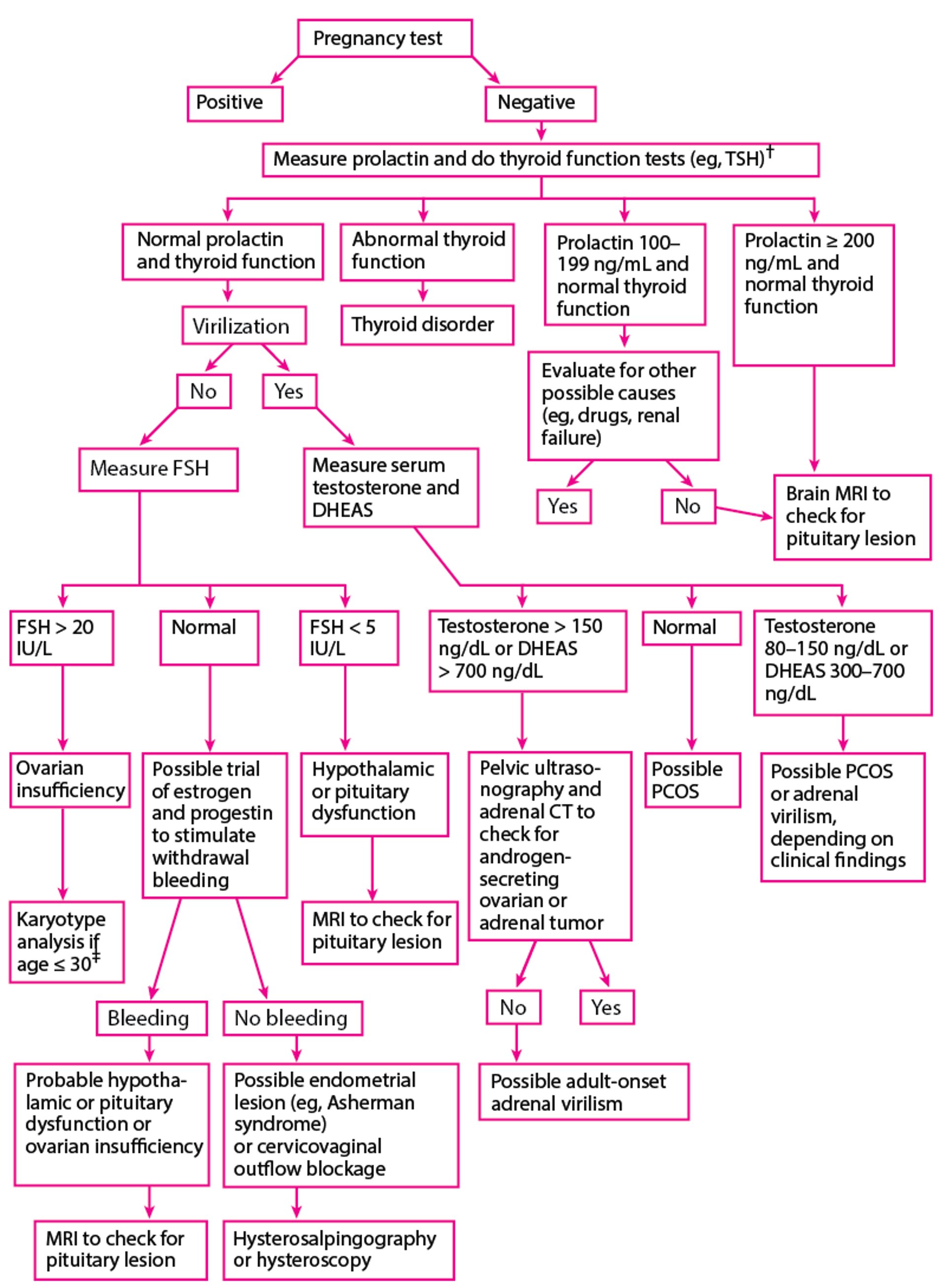
Additional blood tests that are commonly done (to exclude specific etiologies) include:
Follicle-stimulating hormone (ovarian insufficiency); if the level is high, it should be remeasured monthly at least twice
Thyroid stimulating hormone (thyroid disease)
Prolactin (if the level is high [hyperprolactinemia], it should be remeasured)
Total serum testosterone or dehydroepiandrosterone sulfate (PCOS or other causes of hirsutism or virilization)
Amenorrhea with high follicle-stimulating hormone (FSH) levels (hypergonadotropic hypogonadism) suggests ovarian dysfunction. Amenorrhea with low FSH and LH and normal TSH levels (hypogonadotropic hypogonadism) suggests hypothalamic or pituitary dysfunction.
Mildly elevated levels of testosterone or DHEAS suggest PCOS, but levels can be elevated in women with hypothalamic or pituitary dysfunction and are sometimes normal in hirsute women with PCOS. The cause of elevated levels can sometimes be determined by measuring serum luteinizing hormone (LH). In polycystic ovary syndrome, circulating LH levels are often increased, increasing the ratio of LH to FSH.
If symptoms or signs suggest an underlying disorder, specific tests may be indicated. For example, patients with abdominal striae, moon facies, a buffalo hump, truncal obesity, and thin extremities should be tested for Cushing syndrome. Patients with headaches and visual field defects or evidence of pituitary dysfunction require brain MRI. If clinical evaluation suggests a chronic disease, liver and kidney function tests are done, and erythrocyte sedimentation rate (ESR) is determined.
Progestin challenge test
If patients have secondary amenorrhea with normal prolactin and FSH levels and normal thyroid function and do not have virilization, a trial of a progestin can be given to try to evaluate estrogen status. If the estrogen level is sufficient, a course of a progestin should stimulate withdrawal bleeding after the progestin is stopped (progestin challenge test; also called progestin withdrawal test).
The progestin challenge test begins by giving medroxyprogesterone 5 to 10 mg orally once a day or another progestogen for 7 to 10 days. After the last dose,The progestin challenge test begins by giving medroxyprogesterone 5 to 10 mg orally once a day or another progestogen for 7 to 10 days. After the last dose,
If bleeding occurs within a few days, the estrogen level is sufficient and amenorrhea is probably caused by hypothalamic-pituitary dysfunction, ovarian insufficiency, or estrogen excess.
If bleeding does not occur, an estrogen/progestin challenge test is done.
Estrogen/progestin challenge test
The estrogen/progestin challenge test is done by giving an estrogen (eg, conjugated equine estrogen 1.25 mg, estradiol 2 mg) orally once a day for 21 days, followed by medroxyprogesterone 10 mg orally once a day or another progestogen for 7 to 10 days. After the last dose of the progestin, if bleeding does not occur, patients may have an endometrial lesion (eg, Asherman syndrome) or outflow tract obstruction (eg, cervical stenosis). /progestin challenge test is done by giving an estrogen (eg, conjugated equine estrogen 1.25 mg, estradiol 2 mg) orally once a day for 21 days, followed by medroxyprogesterone 10 mg orally once a day or another progestogen for 7 to 10 days. After the last dose of the progestin, if bleeding does not occur, patients may have an endometrial lesion (eg, Asherman syndrome) or outflow tract obstruction (eg, cervical stenosis).
However, bleeding may not occur in patients who do not have these abnormalities because the uterus is insensitive to estrogen due to prolonged use of estrogen/progestin contraceptives or rare endocrine disorders (estrogen insensitivity syndrome, estrogen resistance). Thus, the trial using estrogen and progestin may be repeated for confirmation.
Because this trial takes weeks and results can be inaccurate, diagnosis of some serious disorders may be delayed significantly; thus, brain MRI should be considered before or during the trial if a pituitary or other brain lesion is suspected.
Evaluation references
1. Rebar R. Evaluation of amenorrhea, anovulation, and abnormal bleeding [updated, 2018]. In Endotext [Internet], edited by KR Feingold, B Anawalt, A Boyce, et al. South Dartmouth (MA), MDText.com Inc, 2000. Available from: https://www.ncbi.nlm.nih.gov/books/NBK279144/
2. Munro MG, Critchley HOD, Fraser IS; FIGO Menstrual Disorders Committee. The two FIGO systems for normal and abnormal uterine bleeding symptoms and classification of causes of abnormal uterine bleeding in the reproductive years: 2018 revisions [published correction appears in Int J Gynaecol Obstet. 2019 Feb;144(2):237. doi: 10.1002/ijgo.12709.]. Int J Gynaecol Obstet. 2018;143(3):393-408. doi:10.1002/ijgo.12666
Treatment of Amenorrhea
Treatment is directed at the underlying disorder. Some abnormalities obstructing the genital outflow tract can be surgically repaired.
If a Y chromosome is present, bilateral oophorectomy is recommended because risk of ovarian germ cell cancer is increased.
Common problems associated with amenorrhea may also require treatment, including:
For infertility if pregnancy is desired, inducing ovulation
Treating symptoms and long-term effects of estrogen deficiency (eg, osteoporosis, cardiovascular disorders, vaginal atrophy)
Treating symptoms and managing long-term effects of estrogen excess (eg, prolonged bleeding, persistent or marked breast tenderness, risk of endometrial hyperplasia and cancer)
Minimizing hirsutism and long-term effects of androgen excess (eg, cardiovascular disorders, hypertension)
Guidelines for Amenorrhea
The following is a list of professional medical society or government clinical practice guidelines regarding this medical issue (this is not a comprehensive list):
American College of Obstetricians and Gynecologists (ACOG) Committee Opinion. Management of Acute Obstructive Uterovaginal Anomalies. 2019 (reaffirmed 2021).
ACOG Committee Opinion. Menstruation in Girls and Adolescents: Using the Menstrual Cycle as a Vital Sign. 2015 (reaffirmed 2021).
ACOG Committee Opinion. Müllerian Agenesis: Diagnosis, Management, and Treatment. 2018 (reaffirmed 2024).
ACOG Committee Opinion. Primary Ovarian Insufficiency in Adolescents and Young Women. 2014 (reaffirmed 2021).
Key Points
Amenorrhea is the absence of menses. Primary amenorrhea is failure of menses to occur by age 15 years in patients with normal growth and secondary sexual characteristics. Secondary amenorrhea is absence of menses for 3 months in patients with previously regular menstrual cycles or for ≥ 6 months in patients with previously irregular menses.
Primary amenorrhea may be caused by gonadal dysgenesis, congenital reproductive tract anomalies, or endocrinologic abnormalities.
Secondary amenorrhea may be caused by acquired reproductive tract structural abnormalities that interfere with menstrual function or obstruct menstrual flow or endocrinologic abnormalities.
Evaluate with history, physical examination, and hormonal blood tests (beta-human chorionic gonadotropin, follicle-stimulating hormone, thyroid stimulating hormone, prolactin, and total serum testosterone or dehydroepiandrosterone sulfate). Imaging with pelvic ultrasound or MRI is required if structural abnormalities are suspected. Genetic testing is done if genetic or chromosomal abnormalities are suspected.
If patients have primary amenorrhea and normal secondary sexual characteristics, begin testing with pelvic ultrasound to check for congenital anatomic genital tract obstruction.
If patients have signs of virilization, check for conditions that cause androgen excess (eg, polycystic ovary syndrome, an androgen-secreting tumor, Cushing syndrome, use of certain medications).
If patients have symptoms and signs of estrogen deficiency (eg, hot flashes, night sweats, vaginal dryness or atrophy), check for primary ovarian insufficiency and conditions that cause functional hypothalamic amenorrhea.
If patients have galactorrhea, check for conditions that cause hyperprolactinemia (eg, pituitary dysfunction, use of certain medications).
Drug Information for the Topic


