

Intestinal obstruction is significant mechanical impairment or complete arrest of the passage of contents through the intestine due to pathology that causes blockage of the bowel. Symptoms include cramping pain, vomiting, obstipation, and lack of flatus. Diagnosis is clinical and confirmed by abdominal radiographs. Treatment is fluid resuscitation, nasogastric suction, and, in most cases of complete obstruction, surgery.
(See also Acute Abdominal Pain.)
Mechanical obstruction is divided into obstruction of the small bowel (including the duodenum) and obstruction of the large bowel. Obstruction may be partial or complete. Approximately 85% of partial small-bowel obstructions resolve with nonoperative treatment (1), whereas most complete small-bowel obstructions require surgery.
Reference
1. ten Broek RPG, Krielen P, Di Saverio S, et al. Bologna guidelines for diagnosis and management of adhesive small bowel obstruction (ASBO): 2017 update of the evidence-based guidelines from the world society of emergency surgery ASBO working group. World J Emerg Surg. 2018;13:24. Published 2018 Jun 19. doi:10.1186/s13017-018-0185-2
Etiology of Intestinal Obstruction
Overall, the most common causes of mechanical obstruction are adhesions, hernias, and tumors.
Other general causes are diverticulitis, foreign bodies (including gallstones), volvulus (twisting of bowel on its mesentery), intussusception (telescoping of one segment of bowel into another), and fecal impaction.
Specific segments of the intestine are affected differently (see table ).
Causes of Intestinal Obstruction
Location | Cause |
|---|---|
Colon | Tumors (usually in left colon), diverticulitis (usually in sigmoid), volvulus of sigmoid or cecum, fecal impaction, Hirschsprung disease, Crohn disease |
Duodenum | |
| Cancer of the duodenum or cancer of the head of pancreas, ulcer disease |
| Atresia, volvulus, bands, annular pancreas |
Jejunum and ileum | |
| Hernias, adhesions (common), tumors, Meckel diverticulum, Crohn disease (uncommon), Ascaris infestation, midgut volvulus, intussusception by tumor (rare), foreign body, gallstones (rare) |
| Meconium ileus, volvulus of a malrotated gut, atresia, intussusception |
Pathophysiology of Intestinal Obstruction
In simple mechanical obstruction, blockage occurs without vascular compromise. Ingested fluid and food, digestive secretions, and gas accumulate above the obstruction. The proximal bowel distends, and the distal segment collapses. The normal secretory and absorptive functions of the mucosa are depressed, and the bowel wall becomes edematous and congested. Severe intestinal distention is self-perpetuating and progressive, intensifying the peristaltic and secretory derangements and increasing the risks of dehydration and progression to strangulating obstruction.
Strangulating obstruction is obstruction with compromised blood flow; it occurs in nearly 25% of patients with small-bowel obstruction (1). It is usually associated with hernia, volvulus, and intussusception. Strangulating obstruction can progress to infarction and gangrene in as little as 6 hours. Venous obstruction occurs first, followed by arterial occlusion, resulting in rapid ischemia of the bowel wall. The ischemic bowel becomes edematous and infarcts, leading to gangrene and perforation. In large-bowel obstruction, strangulation is rare (except with volvulus).
Perforation may occur in an ischemic segment (typically small bowel) or when marked dilation occurs. The risk is high if the cecum is dilated to a diameter ≥ 13 cm. Perforation of a tumor or a diverticulum may also occur at the obstruction site.
Pearls & Pitfalls
|
Pathophysiology reference
1. Jancelewicz T, Vu LT, Shawo AE, Yeh B, Gasper WJ, Harris HW. Predicting strangulated small bowel obstruction: an old problem revisited. J Gastrointest Surg. 2009;13(1):93-99. doi:10.1007/s11605-008-0610-z
Symptoms and Signs of Intestinal Obstruction
Obstruction of the small bowel causes symptoms shortly after onset: abdominal cramps centered around the umbilicus or in the epigastrium, vomiting, and—in patients with complete obstruction—obstipation. Patients with partial obstruction may develop diarrhea. Severe, steady pain suggests that strangulation has occurred.
In the absence of strangulation, the abdomen is not tender. Hyperactive, high-pitched peristalsis with rushes coinciding with cramps is typical. Sometimes, dilated loops of bowel are palpable. With infarction, the abdomen becomes tender and auscultation reveals a silent abdomen or minimal peristalsis. Shock and oliguria are serious signs that indicate either late simple obstruction or strangulation.

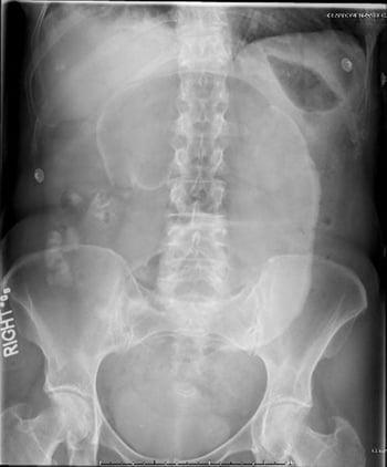
In this abdominal radiograph, the cecum has twisted around its mesentery, causing a dilated "coffee bean" to project toward the left upper quadrant.
Obstruction of the large bowel usually causes milder symptoms that develop more gradually than those caused by small-bowel obstruction. Increasing constipation leads to obstipation and abdominal distention. Vomiting may occur (usually several hours after onset of other symptoms) but is not common. Lower abdominal cramps unproductive of feces occur. Systemic symptoms are relatively mild, and fluid and electrolyte deficits are uncommon.
Physical examination typically shows a distended abdomen with loud borborygmi. There is no tenderness, and the rectum is usually empty. A mass corresponding to the site of an obstructing tumor may be palpable.
Volvulus often has an abrupt onset. Pain is continuous, sometimes with superimposed waves of colicky pain.

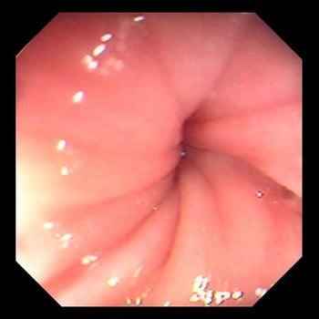
Volvulus is a twisting of the colon around itself, sometimes causing strangulation with ischemia and necrosis. Occasionally, the rotation can be reduced noninvasively with an endoscope. This image shows an endoscopic view of a sigmoid volvulus; twisting of the colonic folds is visible.
Diagnosis of Intestinal Obstruction
Abdominal series

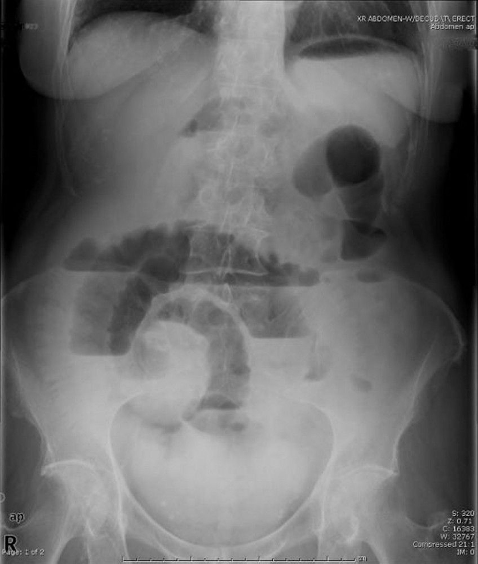
This upright abdominal radiograph shows obstruction of the small bowel. Note the multiple air-fluid levels.
Image provided by Parswa Ansari, MD.
Supine and upright abdominal radiographs should be taken and are usually adequate to diagnose obstruction. Although only laparotomy can definitively diagnose strangulation, careful serial clinical examination may provide early warning. Elevated white blood cells and acidosis may indicate that strangulation has already occurred, but these signs may be absent if the venous outflow from the strangulated loop of bowel is decreased.
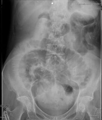
This supine abdominal radiograph shows obstruction of the small bowel. Note the dilated loops of small bowel.
On plain radiographs, a ladderlike series of distended small-bowel loops is typical of small-bowel obstruction but may also occur with obstruction of the right colon. Fluid levels in the bowel can be seen in upright views. Similar, although perhaps less dramatic, radiographic findings and symptoms occur in ileus (paralysis of the intestine without obstruction); differentiation can be difficult. Distended loops and fluid levels may be absent with an obstruction of the proximal jejunum or with closed-loop strangulating obstructions (as may occur with volvulus). Infarcted bowel may produce a mass effect on radiograph. Gas in the bowel wall (pneumatosis intestinalis) indicates gangrene.
In large-bowel obstruction, abdominal radiograph shows distention of the colon proximal to the obstruction. In cecal volvulus, there may be a large gas bubble in the mid-abdomen or left upper quadrant. With both cecal and sigmoid volvulus, a contrast enema shows the site of obstruction by a typical “bird-beak” deformity at the site of the twist; the procedure may actually reduce a sigmoid volvulus. If contrast enema is not done, colonoscopy can be used to decompress a sigmoid volvulus but rarely works with a cecal volvulus.
Abdominal CT is being used more often in suspected small-bowel obstruction.
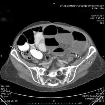
In this CT scan, the small bowel is dilated and filled with air and fluid. Oral contrast is seen in some of the loops but has not traveled down to the distal small bowel. Note the collapsed cecum and sigmoid colon (arrows). A specific point of obstruction (transition point) cannot always be seen on CT, but the dilated proximal bowel and collapsed distal bowel are highly suggestive of the diagnosis.
Treatment of Intestinal Obstruction
Nasogastric suction
IV fluids
IV antibiotics if bowel ischemia suspected
Patients with possible intestinal obstruction should be hospitalized. Treatment of acute intestinal obstruction must proceed simultaneously with diagnosis. A surgeon should always be involved.
Supportive care is similar for small- and large-bowel obstruction: nasogastric suction, IV fluids (0.9% saline or lactated Ringer’s solution for intravascular volume repletion), and a urinary catheter to monitor fluid output. Electrolyte replacement should be guided by test results, but, in cases of repeated vomiting, serum sodium and potassium are likely to be depleted.
If bowel ischemia or infarction is suspected, antibiotics should be given (eg, a third-generation cephalosporin, such as cefotetan or cefoxitin) before operative exploration.If bowel ischemia or infarction is suspected, antibiotics should be given (eg, a third-generation cephalosporin, such as cefotetan or cefoxitin) before operative exploration.
Specific measures
Obstruction of the duodenum in adults is treated by resection or, if the lesion cannot be removed, palliative gastrojejunostomy (for treatment in children, see Duodenal Obstruction).
Complete obstruction of the small bowel is preferentially treated with early laparotomy, although surgery can be delayed 2 or 3 hours to improve fluid status and urine output in a very ill, dehydrated patient. The obstructing lesion is removed whenever possible. If a gallstone is the cause of obstruction, it is removed through an enterotomy, and cholecystectomy need not be done. Procedures to prevent recurrence should be done, including repair of hernias, removal of foreign bodies, and lysis of the offending adhesions.
In most patients with early postoperative obstruction or repeated obstruction caused by adhesions, nasogastric decompression through a sump tube may be attempted in the absence of peritoneal signs. Failure of the obstruction to resolve or development of symptoms or signs of ischemia are indications to proceed with surgery.
Disseminated intraperitoneal cancer obstructing the small bowel is a major cause of death in adult patients with gastrointestinal tract cancer. Bypassing the obstruction, either surgically or with endoscopically placed stents, may palliate symptoms briefly. A palliative venting gastrostomy may also be placed endoscopically.
Obstructing colon cancers can sometimes be treated by a single-stage resection and anastomosis, with or without a temporary colostomy or ileostomy. When this procedure is not possible, a diverting colostomy with delayed resection is recommended. Occasionally, the tumor may be resected and a colostomy or ileostomy is created; the stoma may possibly be closed at a later time. However this approach is sometimes associated with poorer oncologic outcomes. The use of an endoscopic stent to temporarily relieve the obstruction is controversial. Although stenting plays a role in palliation of a left-sided obstructing cancer in patients who may not tolerate an operation, there is potential for perforation, and some studies have suggested a decreased survival rate compared to elective surgical resection when a stent is used to bridge a potentially curable obstructing cancer.
When diverticulitis causes obstruction, perforation is often present. Removal of the involved area may be very difficult but is indicated if perforation and general peritonitis are present. Resection and colostomy are done, and anastomosis is often postponed.
Fecal impaction usually occurs in the rectum and can be removed digitally and with enemas. However, a fecal concretion alone or in a mixture (ie, with barium or antacids) that causes complete obstruction (usually in the sigmoid) requires laparotomy.
Treatment of cecal volvulus consists of resection and anastomosis of the involved segment or fixation of the cecum in its normal position by cecostomy in a patient who is frail. In sigmoid volvulus, an endoscope or a long rectal tube can often decompress the loop, and resection and anastomosis may be deferred for a few days. Without a resection, recurrence is almost inevitable.
Key Points
The most common causes of obstruction are adhesions, hernias, and tumors; a small-bowel obstruction in the absence of prior surgery or hernias is often caused by a tumor.
Vomiting and third spacing of fluid cause volume depletion.
Prolonged obstruction can cause bowel ischemia, infarction, and perforation.
Use nasogastric suction and IV fluids before surgical repair.
Consider a trial of nasogastric suction rather than immediate surgery for patients with recurrent obstruction due to adhesions.
Drug Information for the Topic


